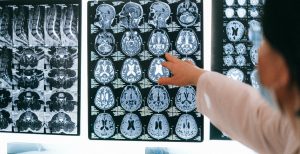
Researchers at the Royal Adelaide Hospital (RAH) are investigating the next generation tumour imaging technique to better equip oncologists in the fight against fatal glioblastoma brain tumours.
The FET-PET in Glioblastoma (FIG) study aims to compare traditional MRI imaging with the newer FET-PET at both diagnosis as well as in measuring the effects of treatment.
Room for improvement
Glioblastoma is the most common and aggressive malignant brain tumour in adults, affecting approximately 1000 Australians annually.
It remains an incurable tumour with a median survival of only 15 months and affects young people in the prime of life as well as the elderly.
Despite advances in therapy, there have been no significant reductions in incidence and mortality rates in the last decade.
“Glioblastoma is a disease that is very difficult to manage and it is important we have as many tools as possible available,” said Associate Professor Hien Le, lead investigator of the FIG study at the RAH.
“We can make the biggest gains in diseases where prognosis is poor and research gaps exist. That’s why we’re really keen to bring on the TROG 18.06 FIG study, because anything that shows a benefit is going to go a long way.”
MRI versus FET-PET
Imaging provides initial information to assist diagnosis, guides surgical removal of the tumour and radiotherapy planning, and is used to monitor treatment response.
Currently, almost all of this information is obtained from MRI, and however valuable this information is, there are areas of active tumour that may be missed by MRI.
Additionally, following treatment, MRI is limited in its ability to distinguish between progression of the disease and damage to normal tissue from the treatment itself. This uncertainty can cause anguish to patients and families while waiting for more data with which to make the distinction and can lead to the inappropriate cessation of effective therapy.
A newer form of imaging has been developed using Positron Emission Tomography (PET), where a radiotracer called FET, a chemical compound injected into the bloodstream, is used to detect whether tumour cells are active or not.
Compared to MRI, FET-PET promises to more accurately define the tumour and treatment areas for radiotherapy planning, and more accurately assess tumour changes versus treatment-related changes. The FIG study aims to test these supposed advantages by comparing the two imaging techniques both prior to treatment and following treatment.
In the first part of the study, radiation oncologists will use MRI to examine tumours of patients prior to radiation therapy, establishing the size of the tumour and treatment target. They will then do the same thing using FET-PET and compare the sizes determined using the two techniques.
Similarly, in the second part of the study, radiation oncologists will compare the observations using MRI and FET-PET of patients who have undergone radiation therapy.
A changing landscape for glioblastoma
“FET-PET is an investigative tool that is not yet supported by Medicare. If the FIG study demonstrates an advantage to using FET-PET in this clinical setting, then that will go a long way in terms of lobbying the federal government which will therefore influence how we manage patients due to improved access,” said A/Prof Le.
“It’s data that will potentially allow us as clinicians to systematically insert FET-PET into the treatment landscape for glioblastoma.”
The FIG trial is supported by Trans Tasman Radiation Oncology Group (TROG) and started in late 2021. It will continue for the next two or three years, targeting the involvement of 210 participants.
“The RAH is the largest academic health institution in the state,” said A/Prof Le.
“I think it’s vital for us to be involved in trials like this, that are asking really important questions that may impact healthcare for a critical disease like glioblastoma.”
“It is not easy running trials like this, but it is worth it. It’s where we can make a big impact.”


